Stem Cells May Hold the Key to Helping Patients with Cleft Palate
Posted
09 Oct 17
Ostrow Professor and Associate Dean of Research Yang Chai’s research is heavily focused on cleft lip and cleft palate and how stem cell therapy might be used to detect — and even prevent — cleft formation.
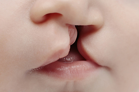
Every time we drink or breathe, the soft palate is hard at work. It acts like a trap door, allowing air to squeeze through or shutting so liquids can pass by. And when congenital problems arise there, like a cleft in the palate, they can cause problems with speech, swallowing, breathing and hearing.
Yang Chai’s work offers a genetic and evolutionary view of cleft palate — and suggests new treatments may help patients in the future.
“We as humans have a very unique soft palate,” explains Chai DDS ’91 PhD ’96. The reason? Our ability to talk. In many other animals, the structure is fused to the posterior pharyngeal wall. But humans have a flexible structure, where air can be pushed and squeezed through. This so-called oropharyngeal region also changes over a person’s life: babies can suck milk and breathe at the same time, but after early life, people lose that ability.
Chai’s lab is particularly interested in finding the cause of cleft lip and cleft palate. As he explains, these problems are one of the most common congenital birth defects — occurring in 1 in 1,000 live births, second only to congenital heart defects. They can also be devastating. On average, each patient with cleft palate has multiple surgical procedures from birth to adulthood to close the cleft, including bone grafts, implants and orthodontic work.
Despite the complexity of this area, the soft palate hasn’t been studied too deeply, because it’s difficult to find animal models.
Discovery could lead to fewer surgeries
In a new study published in the journal Development, Chai and his colleagues used engineered mice to zero-in on how the oropharyngeal region is patterned, and how a special group of cells — cranial neural crest cells — come in and help set up scaffolds for muscle cells during craniofacial development.
“From this study, we now have a better understanding of how we as humans have a palate that is different from other animals,” says Chai, director of USC’s Center for Craniofacial Molecular Biology.
Looking to the future, Chai’s work suggests that knowing how cells develop scaffolds in this area can help reduce the amount of surgeries it takes to close cleft palates.
When it comes to surgical procedures, one of the challenges craniofacial surgeons face is they often don’t have sufficient muscle — or they can’t lay the muscle fibers in the right direction.
“The hope is that we can regenerate muscle and that can be used to help improve the surgical correction of a cleft in the soft palate,” Chai explains. “From this study, we have learned that it actually has to be another cell type — cranial neural crest cells — that have to be there to give the right signal for the muscle to go in the right place.”
Chai is also hoping to raise awareness about clefts — particularly a specific type of submucosal cleft — that don’t often get diagnosed until a child is far into toddlerhood. “In a lot of these cases, the kids are not diagnosed early enough to receive proper treatment, simply because the parents don’t see a gap in their mouth,” he explains.
It’s only when the kid begins to learn to talk that parents realize their kids are not speaking in a normal way. Chai encourages pediatricians to examine the newborns and look at their soft palate during a cry to make sure there isn’t a cleft there.