Scanner Gives a GPS Map to Endodontists
Posted
10 Jan 23
The new tech allows endodontists to see in three dimensions, allowing more accurate diagnoses, treatment and evaluation.
ENDODONTISTS SPECIALIZE IN SAVING NATURAL DENTITION, mostly focusing on the tooth’s interior. They live and die by imaging, both to diagnose what’s going on inside a tooth as well as to see how a treatment is working.
Historically, that’s been done with traditional two-dimensional radiographs, which offer a flat view of a tooth.
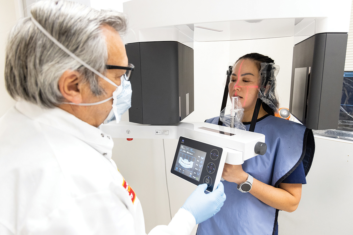
Recently, Ostrow received a cone beam computed tomography scanner for use in the advanced education program in endodontics, thanks to a generous donation from endodontist members of the Friends of Dentistry, particularly Samir Batniji ENDO ’87.
The scanner rotates once around a patient’s head, giving a crisp 3-D view of what is going on inside teeth that are basically embedded in bone. It opens up new possibilities of seeing inside teeth, before a procedure ever starts.
Takes the Guesswork Out
“I call it GPS for teeth,” said Rafael Roges, associate professor of clinical dentistry and director of the advanced education program in endodontics. “It’s like a 3-D map of a tooth’s anatomy, and it really is changing what we do.” The school has done 815 scans since June 2022, he said, at the time of publication.
Roges said the technology gives endodontists a better idea of what is going on inside and around the roots of the tooth before they start working there, making it a far better way than traditional radiographs to diagnose, treat and evaluate patients. The technology was developed in the mid-1990s and has been used in dentistry since the early 2000s.
Endodontics was blind, but now we see. We can’t live without [the scanning technology] anymore.
—Rafael Roges, Director of the Advanced Education Program in Endodontics
For Mattison Donaldson ’15, DDS ’20, ENDO ’23, the scanner allowed her to diagnose a patient who could have been overlooked otherwise. Donaldson said she took traditional mandibular radiographs, and what she found looked healthy and normal. But then she took a scan, and found a huge lesion — revealing that the existing root canal was not healing and needed re-treatment. “You see so much more than you can through other ways,” she said. “It takes the guesswork out of your actual treatment.”
The scanner can also help endodontists plan ahead for surgeries and pinpoint the exact location where they need to work, so they don’t end up removing bone that could otherwise be preserved, said Ariana Malek ENDO ’23. “We can see every aspect of the tooth, and even if it looks simple, we have been getting scans on almost every case,” she said. “With it, the surgeries go smoother.”
Once Was Blind But Now Can See
The scanner is safe to use, Roges said. Having a three-dimensional view often changes a diagnosis: In a study of cone beam computed tomography versus two-dimensional digital radiography, researchers found that having the technology changed a patient’s diagnosis 62 percent of the time.
“It’s becoming the best way to diagnose, treat and evaluate what we do,” he said, adding that the additional cost is mitigated by better treatments that don’t need to be redone. The scanner is mostly used for complicated anatomy, trauma or root canals that need to be redone — but, in Roges’ opinion, it should be used for everyone. “Endodontics was blind, but now we can see,” he said. “We can’t live without it anymore.”
Recent news
A new clinic for treating precancerous oral lesions using several treatment options, including the first prospective clinical trial for imiquimod — a medication approved for skin cancer. It could revolutionize oral cancer treatment.