Ostrow researcher Yang Chai’s scientific discovery could lead to effective biological treatment for a common birth defect
Posted
23 Mar 15
Research findings presented in article published in April 2015 issue of Nature Cell Biology
Throughout every human and animal’s body, stem cell populations are responsible for the growth, regeneration and repair of tissues. While the power of some types of stem cells is already being used in cutting-edge medicine, there is still much to discover before we truly unlock their potential.
Ostrow researchers have discovered which stem cells are responsible for the growth of craniofacial bones in mice—a finding that could have a profound impact on the understanding and treatment of craniosynostosis, a birth defect that can lead to an array of physical and intellectual disabilities in humans.
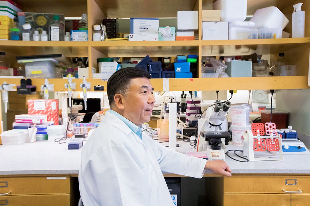
In an article published in the April 2015 issue of Nature Cell Biology, Professor Yang Chai, holder of the George and Mary Lou Boone Chair of Craniofacial Biology, describes how he and postdoctoral fellow Hu Zhao identified a population of Gli1+ stem cells within the mesenchyme of tissue between the bones of the skull, known as cranial sutures.
Chai’s research team noted that if Gli1+ cells were eliminated from the sutures, the craniofacial bones experienced growth arrest and weakening, leading researchers to conclude these cells were indispensable in healthy skull development.
The findings could have a significant impact on the treatment of craniosynostosis, a birth defect affecting one in 2,500 births.
In a normally developing baby, the craniofacial bones do not grow together for up to 18 months to allow for the brain to reach nearly 70 percent of its eventual size. It’s these open sutures that cause babies’ heads to have “soft spots.”
In an infant with craniosynostosis, the skull bones fuse together before the brain has fully developed, which can cause an array of problems, including hearing and vision defects and developmental disorders.
“Traditional scientific literature viewed craniosynostosis as an overgrowth of bone,” Chai said. “Our findings offer a brand new perspective, which is that craniosynostosis could actually be viewed as a lack of stem cells.”
The discovery could lead to a different treatment intervention for babies with craniosynostosis. Up until now, if the bones prematurely fused, a doctor had to operate, typically within the first year of the baby’s life. The surgery entails removing the top of the child’s skull, cutting it into segments and then stapling it back together, creating enough space for the infant’s developing brain to grow.
Thanks to the discovery of Gli1+ stem cells, this mechanical intervention could soon be replaced with a biological intervention—something Chai and his team are already working on.
Chai is the Associate Dean of Research at the Herman Ostrow School of Dentistry of USC and the Director of the Center for Craniofacial Molecular Biology.
He earned his doctor of dental medicine degree from Peking University in China in 1984, before moving to USC to pursue doctor of philosophy and doctor of dental surgery degrees.
He is widely recognized throughout the dental and craniofacial research community for his investigations of the molecular and cellular mechanisms of craniofacial development, including oral and facial birth defects such as cleft palate.
“I was trained in oral and maxillofacial surgery so I operated a lot on kids with congenital birth defects,” he said. “We always wanted to understand more about what causes these congenital malformations with the hopes to provide a better diagnosis and possible prevent them in the future.”
This study was supported by grants from the National Institute of Dental and Craniofacial Research, NIH (DE022503, DE020065 and DE012711) to Yang Chai.
Related:
Read Dr. Chai’s Nature Cell Biology article