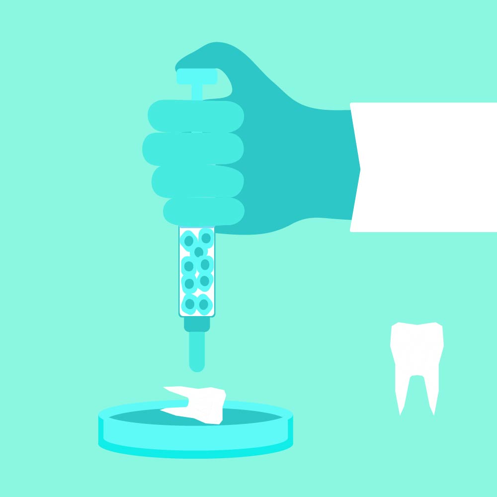
Figuring out how teeth are built, one cell at a time
Posted
13 Sep 22
USC researchers look inside teeth to figure out how we might regenerate teeth in the future.
TEETH ARE MARVELOUSLY COMPLICATED structures — and the way they develop is also complex.
The majority of tooth tissue (except the enamel) comes from cranial neural crest cells — stem cells that eventually develop into craniofacial bones and cartilage. When these cells develop abnormally, it can lead to birth defects including cleft lip, cleft palate and tooth malformation.
USC scientists have been trying to understand how these cells “decide” which type of cell they will become — for example, the hard part of the tooth known as dentin, or a ligament that supports a tooth.
“If we know how these cells are choreographed in the development process, then we can have a better understanding about how certain genetic mutations may affect tooth development,” Associate Dean of Research Yang Chai PhD ’91, DDS ’96 said. “This information will become a kind of a blueprint that will guide us to build a certain part of tooth structure, or even to regenerate parts of the tooth in the future.”
The ways that cranial neural crest cells form diverse tissues have remained largely unknown. In a recently published study in Nature Communications, Chai and his colleagues were able to use mouse molar development as a model to build an atlas showing how individual cells made their decision through space and time. It’s the first time research has shown how particular cellular domains interact with each other at the single cell resolution to form the dental tissues and identified specific molecules and signaling pathways that regulate dental development.
Chai said the discovery allows researchers to go deeper — like moving from a snapshot of a football game to a move, where you can see each player changing location in space over time. “Now we know for each cell, what type of genes they express, how they position themselves,” he said, “and more importantly, where they go when they begin to form parts of the tooth.”
The next steps are to move the work into a larger animal model — and eventually into tooth regeneration in humans. They hope this work can be used in the future to provide better treatments for patients. Think about someone with a dental implant: that structure gets fused into the jaw bone — as opposed to a natural tooth, which is held in place by ligaments. When a person bites down, a tooth gives feedback: it needs a different force to crush an ice cube versus a piece of cake.
“When you chew those foods, you know, there’s some kind of natural feedback between the tooth and the periodontal ligaments, and there are nerve endings, but for implants, that’s not the case because they are fused to the bone,” Chai said. He hopes to use the information from this study as part of the building block towards rebuilding parts of the teeth and ligaments to create better teeth in the future.
“Dental implants are very successful,” he added. “But does that mean this is the best we can do? I think we can do better in tooth regeneration.”
Recent news
A new clinic for treating precancerous oral lesions using several treatment options, including the first prospective clinical trial for imiquimod — a medication approved for skin cancer. It could revolutionize oral cancer treatment.